Omental Bursa
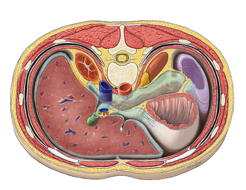
A cross-sectional anatomical illustration in textbook style
When learning human anatomy, students usually find it hard to understand the peritoneal relationship between abdominal viscera. The structures around the omental foramen and omental bursa are extremely complex and they are difficult to identify in dissection labs.
This illustration elucidates the structural complexity surrounding the omental bursa by providing a cross-sectional view of the abdomen. Reconstructed from a CT scan at T12 vertebra, this illustration shows the omental bursa is bounded by hepatoduodenal ligament, lesser omentum, stomach, gastrosplenic ligament, splenorenal ligament and the posterior abdominal wall. The peritoneal relationships for five organs are clarified. It demonstrated that liver, stomach and spleen are intraperitoneal, whereas the two kidneys are retroperitoneal.


Faculty advisor
Dave Mazierski and Shelley Wall
Content advisor
Anne Agur
Medium/Software
Procreate and Adobe Illustrator
Final presentation format
Print, 1/2 page in a textbook
Primary audience
Students in human anatomy classes
Work Process
Reference
-
Agur, A. M. R., & Dalley, A. F. (2008). Grant’s Atlas of Anatomy, 12th Edition (Grant, John Charles Boileau//Grant’s Atlas of Anatomy) (12th ed.). Lippincott Williams & Wilkins.
-
Woodburne, R. T. (1978). Essentials of human anatomy (6th ed.). Oxford University Press.
-
Eycleshymer, A. C., & Schoemaker, D. M. (1923). A Cross Section Anatomy. New York, Appleton-Century-Crofts.
-
Netter, F. H. (1997). Atlas of Human Anatomy, 2nd Edition (2nd ed.). Novartis.
-
S. (2001). Atlas of Human Anatomy. Springhouse Pub Co.

